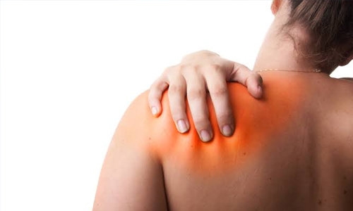
Rotator Cuff Tendinitis
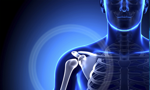
The shoulder is made up of bones, Upper Arm (Humerus), Shoulder Blade (Scapula) and Collar Bone (Clavicle). Arm Is kept in shoulder socket by rotator cuffs. These muscles & tendons form a covering around the head of upper arm bone and attached to shoulder blade. There is a lubricating sac called a bursa between the rotator cuff and the bone on top of shoulder (Acromion). The bursa allows the rotator cuff tendons to glide freely when during movement of the arm.
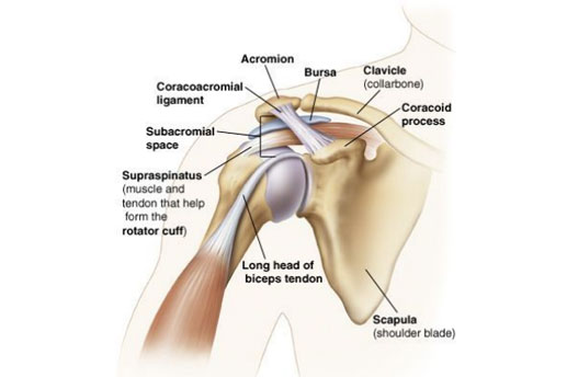
Normal Anatomy of the shoulder
The rotator cuff is a common source of pain in the shoulder. Pain can be the result of Tendinitis in which the rotator cuff tendons can be irritated or damaged. Pain can be due to Bursitis in which the bursa can become inflamed & swell with more fluid causing pain. In Impingement, the space between the acromion & rotator cuff narrows when the arm is raised to the shoulder height. The acromion can rub against (or impinge on) the tendon & the bursa causing irritation and pain.
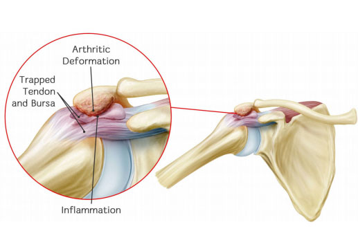
Acromion Impinges on the rotator cuff & bursa
Rotator cuff pain is common in middle-aged people & young athletes who use their arms overhead for swimming, baseball & tennis are particularly vulnerable. Those who do repetitive lifting or overhead activities using the arm are also susceptible. Pain may also develop as the result of a minor injury, sometimes, it occurs with no apparent cause.
Rotator cuff pain causes local swelling & tenderness in the front of the shoulder. There is pain and stiffness may present during arm movement. Beginning symptoms may be mild. During early stage patient may have pain radiating from the front of the shoulder to the side of the arm, also have minor pain that is present both with activity & at rest. As the problem progresses, the symptoms increases and patient have pain at night, loss of strength & motion. Difficulty doing activities that place the arm behind the back, such as buttoning or zippering. If the pain comes on suddenly, the shoulder may be severely tender. All movement may be limited & painful.
Rotator cuff Tendinitis can be diagnosed by a patient’s medical history & physical examination. Imaging test may help in diagnosis. X-rays do not show the soft tissue like the rotator cuff. Plain x-rays of a shoulder with rotator cuff pain are usually normal or may show a small bone spur. A special x-ray view called as “Outlet View” may show small bone spur on the front edge of the acromion. MRI & Ultrasound can show fluid or inflammation in the bursa & rotator cuff. In some cases, partial tearing of the rotator cuff will be seen.
The goal of treatment is to reduce pain and restore function. Initial treatment is nonsurgical treatment. It may take several weeks to months, many patients experience a gradual improvement & return to function. Rest & activity modification advisable. Non-steroidal anti-inflammatory medicines (like ibuprofen & naproxen).
Physical Therapy:A physiotherapist initially focuses on restoring normal motion of shoulder joint. Stretching exercises are very helpful to improve range of motion. If patient has difficulty in reaching behind his back patient may have developed tightness of posterior capsule of the shoulder (Capsule refers to the inner lining of the shoulder & posterior refers to the back of the shoulder). Stretching of the posterior capsule can be very effective in relieving pain in the shoulder. Once pain is improving physiotherapist start strengthening of the rotator cuff muscles.
Steroid Injection:If rest, medications & physical therapy do not relieve pain, an injection of a local anaesthetic & a cortisone preparation may be helpful. (Cortisone is a very effective anti-inflammatory medicine when injected into the bursa beneath the acromion can relieve pain)
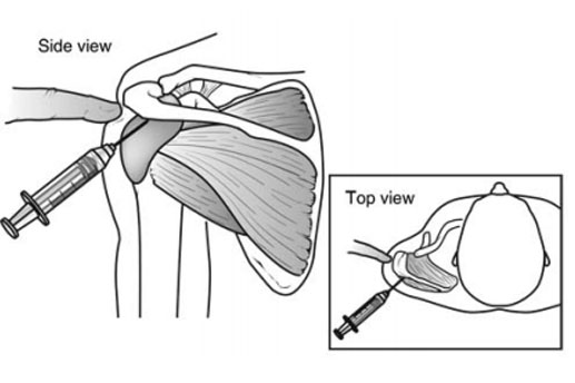
Cortisone injection in shoulder joint
This approach is used when non-surgical treatment does not relieve pain. The goal of surgery is to create more space for the rotator cuff. To do this the doctor will remove the inflated portion of the bursa. A part of the acromion is removed (Anterior acromioplasty). This is also known as a subacromial decompression which is performed using either an arthroscopic or open technique.
Arthroscopic Technique:In this technique, thin surgical instruments are inserted into 2 or 3 small puncture wound around the shoulder. The doctor examines shoulder through a fiberoptic scope connected to a television camera. He/She guides the small instruments using a video monitor & removes bone & soft tissue. In most cases, the front edge of the acromion is removed along with some of the bursal tissue.
Open Surgical Technique:In this technique, the doctor will make a small incision in front of the shoulder. This allows the doctor to see the acromion & rotator cuff directly.
Rehabilitation:After the surgery arm may be placed in a sling for a short period of time. This slows early healing as soon as comfort allows, doctor, will remove the sling to begin exercises and use of the arm. It typically takes 2 to 4 months to achieve complete relief of pain, but it may take up to a year.
The shoulder is made up of bones, Upper Arm (Humerus), Shoulder Blade (Scapula) and Collar Bone (Clavicle). Arm Is kept in shoulder socket by rotator cuffs. These muscles & tendons form a covering around the head of upper arm bone and attached to shoulder blade. There is a lubricating sac called a bursa between the rotator cuff and the bone on top of shoulder (Acromion). The bursa allows the rotator cuff tendons to glide freely when during movement of the arm.
The rotator cuff is a common source of pain in the shoulder. Pain can be the result of Tendinitis in which the rotator cuff tendons can be irritated or damaged. Pain can be due to Bursitis in which the bursa can become inflamed & swell with more fluid causing pain. In Impingement, the space between the acromion & rotator cuff narrows when the arm is raised to the shoulder height. The acromion can rub against (or impinge on) the tendon & the bursa causing irritation and pain.